ENT Surgical Consultants Joliet, New Lenox, Morris

Cholesteatoma

What is a cholesteatoma?

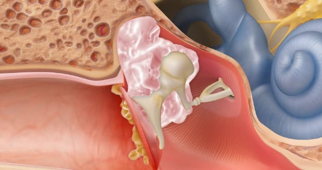
A cholesteatoma is skin growth occurring in the middle ear or mastoid bone behind the eardrum. It is sometimes also called a skin cyst. This is caused by an ingrowth of the skin of the eardrum in most cases.

Why is skin behind my eardrum a problem?

Skin is always regenerating. Once your skin cells die everywhere else in your body, they flake off or exfoliate. If there is skin is in the middle ear, there is nowhere for it to escape as it enlarges. The cholesteatoma growth is slow, but over time skin can erode bone and damage nerves. Cholesteatomas can also become chronically infected, leading to infections and foul-smelling drainage.

How did skin end up in my middle ear?

For most people, it occurs because their eustachian tube is not working properly. Your eustachian tube is supposed to equalize pressure between your middle ear and the outside world. If it is not equalizing pressure the air in your middle ear is absorbed by your body and negative pressure results, leading to your eardrum being sucked in (retracted). Since the outer layer of your eardrum is skin, this can lead to that skin becoming trapped on the wrong side of the eardrum. There are several other less common causes.

CholesteatomaWhat are the normal symptoms from a cholesteatoma?

The most common symptoms are ear drainage, hearing loss, and ear fullness. Sometimes ringing (tinnitus) can occur because of the hearing loss. Infrequently it can cause pain.

Is it cancer?

No. There is no possibility of it becoming cancer.

Is it dangerous?

Cholesteatomas can be dangerous. There are a lot of important structures around the middle ear that could potentially be damaged. Cholesteatomas grow slowly, but if left unchecked can have severe consequences.

Uncommon complications that can occur if left untreated:

  • Vertigo – If skin erodes into the balance canal of the inner ear.
  • Deafness – If skin erodes into the inner ear total deafness can occur.
  • Facial weakness – Your facial nerve runs through your middle ear and can be damaged by cholesteatomas.
  • Cerebrospinal fluid leak – This can occur if the cholesteatoma erodes through the roof of your ear bone (temporal bone), which is also the floor of your cranium (where your brain sits).
  • Meningitis/brain abscess – Your brain is adjacent to your ear and cholesteatoma can potentially lead to severe brain infections.

How are cholesteatomas treated?

In order to remove a cholesteatoma, surgery is necessary. Initially, cholesteatomas often present when they are infected, and they are treated with either oral or topical antibiotics. This can alleviate the initial symptoms, yet surgery is still recommended to avoid damage to critical structures in and around the ear.

A CT scan is usually ordered to determine whether the cholesteatoma has eroded any critical structures, such as the inner ear, facial nerve, brain cavity, and to help with surgical planning. Sometimes an MRI is also needed.

What are the goals of surgery?

  • Priority 1) Safe ear – The highest priority is that the cholesteatoma does not reach your brain, damage your facial nerve, or cause total deafness in that ear.
  • Priority 2) Dry ear – The next priority is to have an ear that does not frequently drain or have infections after surgery.
  • Priority 3) Hearing ear – The last, but still important, priority is to have an ear that hears well following surgery.

All three goals are always sought, however depending on the severity of the cholesteatoma, they cannot always be achieved.

What does the surgery entail?

The surgery is performed under general anesthesia. A small incision is usually made behind the ear, but that can sometimes be avoided if the cholesteatoma is caught early enough. Ear endoscopes are often used to help remove cholesteatoma from hard to reach parts of the middle ear.

There are three possibilities outcomes for the initial surgery:

  1. Ear canal wall left intact, single surgery – The cholesteatoma is isolated, and the surgeon is confident it has completely been removed. Your hearing would then be reconstructed during that surgery and no additional surgery would be needed.
  2. Ear canal wall left intact, second look surgery needed in 6-12 months – The cholesteatoma has been removed, but there is a good chance there are a skin cells left. If only a single skin cell is left in the middle ear, the cholesteatoma will recur. If single cells are left, in 9 months they will grow into pearls which are easily visible and removable in a second look surgery. Hearing would then be reconstructed during the second surgery.
  3. Ear canal wall removal – The back of the ear canal is in the way when removing cholesteatomas. If the cholesteatoma is extensive and the surgeon does not think it can be removed completely with the back of the ear canal intact, it is necessary to remove it with what is called a canal wall down mastoidectomy. This leads to a larger hole for your ear canal, which may be visible to people looking at your ear after surgery. This externalizes the middle ear and mastoid so that skin will come out on its own, so cholesteatoma cannot recur. This cavity will need to be cleaned out periodically by an ENT following this surgery to maintain it. We try to avoid this, but it is sometimes absolutely necessary.
    Usually we cannot say until the surgery is in progress which of these options will be appropriate for you.

What is the recovery from surgery?

You will receive narcotic pain medication just in case, however most patients don’t need it. Tylenol and ibuprofen are usually enough to manage the pain from surgery. Recovery means different things to different people, and everyone bounces back at different rates. In general, after a couple of days of recovery patients are back to their normal routine. You can shower the day after surgery – if there is an incision it is ok to get it wet, but do not scrub directly on it. It helps healing to apply plain Vaseline daily to the incision.

What are the risks of surgery?

  • Anesthesia
  • Bleeding
  • Dizziness
  • Hearing loss – The cholesteatoma itself can often transmit sound itself, so removal can lead to worsened hearing, particularly the hearing bones cannot be reconstructed during the first surgery. There is also a risk of total deafness in the ear following surgery if the cholesteatoma is invading inner ear structures (rare).
  • Recurrence – If a single skin cell is left after the final surgery with the ear canal wall left intact, the cholesteatoma can come back in the future.
  • Facial nerve injury – This is uncommon but possible, particularly in severe cholesteatomas because they can eat away the bone surrounding the facial nerve.
  • Cerebrospinal fluid leak – This can occur if the cholesteatoma erodes away the bone under the brain.

Schedule Your Appointment!

Please call one of our locations: West Joliet 815-725-1191, New Lenox 815-717-8768, and Morris 815-941-1972 to schedule an appointment.

Contact Us Today!

© ENT Surgical Consultants LTD. - All Rights Reserved - Managed by Practis